Der Luebeck EUS Trainer in der Praxis
The Luebeck EUS Trainer in practice
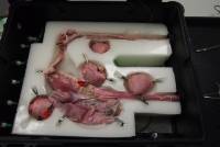
Bild 1: In der Bodenmatrix eingebettete Organe und Punktionsobjekte, die zusätzlich mit Klemmen am Magen bzw. dem Rektum fixiert sind.
Image 1: Imbedded organs and puncture objects into the lower matrix. They are fixed with clips at the stomach or at the rectum.
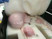
Bild 2: Mit Luft gefüllter Magen und die an der Speiseröhre und dem Magen angelagerten, gefüllten Harnblasen, die als "Pseudozysten" dienen.
Image 2: Air filled stomach and bladders, imitating "pseudocysts" close to the esophagus and the stomach.
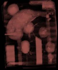
Bild 3: Anordnung der Organe und Punktionsobjekte im CT-Topogramm, da der LET auch für Durchleuchtung und Fusionsbildgebung geeignet ist.
Image 3: Arrangement of the organs and puncture objects in a CT topogram, because the LET is suitable for fluoroscopy and image fusion technology.
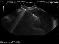
Bild 4: Endosonografisches Abbild einer Punktion einer Pseudozyste mit einer 19 G Nadel durch die Magenwand.
Image 4: endosonographic image of a pseudocyst puncture with a 19 G needle through the stomach wall.
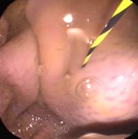
Bild 5: Realitätsgetreue Darstellung des endoskopischen Bildes nach Legen eines Führungsdrahtes in eine Pseudozyste.
Image 5: Realistic depiction of the endoscopic view after the insertion of a guide wire into a pseudocyst.
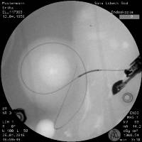
Bild 6: Korrespondierendes Durchleuchtungsbild mit einem in der Pseudozyste aufgerolltem Draht.
Image 6: Corresponding fluoroscopic image with the rolled up guide wire within the pseudocyst.
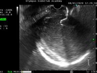
Bild 7: Selbst-expandierender Stent zwischen Pseudozyste und Magenwand.
Image 7: Self-expanding metal stent between pseudocyst and stomach wall.
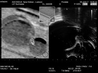
Bild 8: Urologisches Training in Bildfusionstechnik - links CT Schnitt - rechts rektal-sonografisches Bild.
Image 8: Image fusion technology for urological training - left CT scan - right rectal ultrasound.